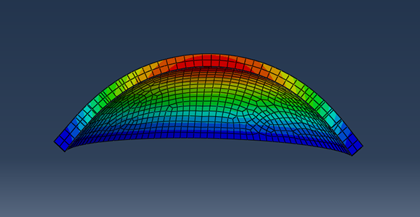
Corneal Biomechanics
The cornea possesses a unique combination of mechanical stiffness, strength, and optical transparency that enables it to serve as both a protective covering and the primary refractive component of the eye. The cornea's contribution to ocular image formation can be degraded by abnormalities in shape, by diseases and the effects of surgery; so an understanding of the biomechanical behavior of the cornea is of great clinical importance to the development of predictive tools to aid the clinical management of patients. The finite element method (FEM) is a computational tool that can be used to represent the geometric, biomechanical, and biological characteristics of a structure. A sample cornea mapped using FEM can be seen in the figure below:
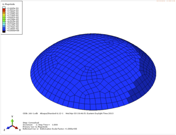
Models have been proposed using the finite element method to model the actual corneal constitutive behavior, and from it, to predict the outcome of corneal refractive surgical procedures. We can develop a patient-specific individualized finite element model for the cornea to compare predicted and in vivo refractive outcomes and to estimate the corneal elastic property changes associated with each procedure by controlling the geometry, mechanical properties and other conditions in the finite element model.